ZEISS | Stemi 508 Greenough Stereomicroscope with 8:1 Zoom
LLS ROWIAK LaserLabSolutions GmbH
Garbsener Landstraße 10
30419 Hannover
Deutschland/Germany
Web: https://www.lls-rowiak.de/
E-Mail: info@lls-rowiak.de
Telefon/Phone: +49 (0)511 277 2976
Fax: +49 (0)511 277 1766
Details
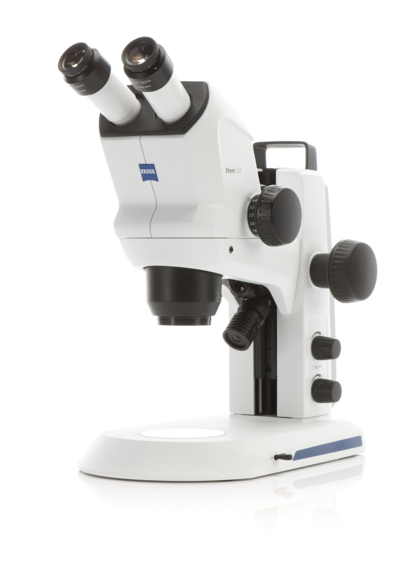
ZEISS Stemi 508 Greenough Stereomicroscope with 8:1 Zoom
Sharp 3D Imaging
Experience contrast-rich, highly accurate 3D images with apochromatic optics and an ergonomic 35° viewing angle. The 8:1 zoom captures minute details in high contrast, ensuring clarity even during prolonged use.
Robust Design for Intense Workloads
Designed for heavy workloads, the Stemi 508 maintains a balanced 3D impression. Whether zooming continuously or using reproducible modes, the image remains sharp and focused throughout the entire magnification range, thanks to its reliable mechanics.
Large Field of View and Apochromatic Correction
Benefit from a sharp, distortion-free, and three-dimensional image with apochromatic zoom optics and efficient stray light suppression. Explore objects in up to 122mm-wide fields of view, and utilize the 8:1 zoom for high-contrast examination of intricate details.
Precision Mechanics Under High Workload
Stemi 508's robust and reliable mechanics are designed for high workloads. Zoom continuously or use the reproducible mode with activated detent positions – the image remains sharp and focused across the entire magnification range. The small 35° viewing angle ensures comfortable work even during extended microscope sessions.
Versatile for All Applications
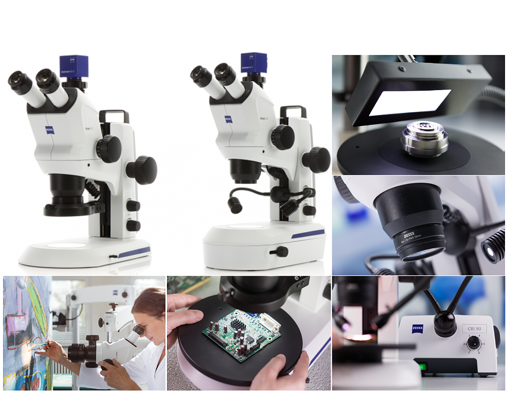
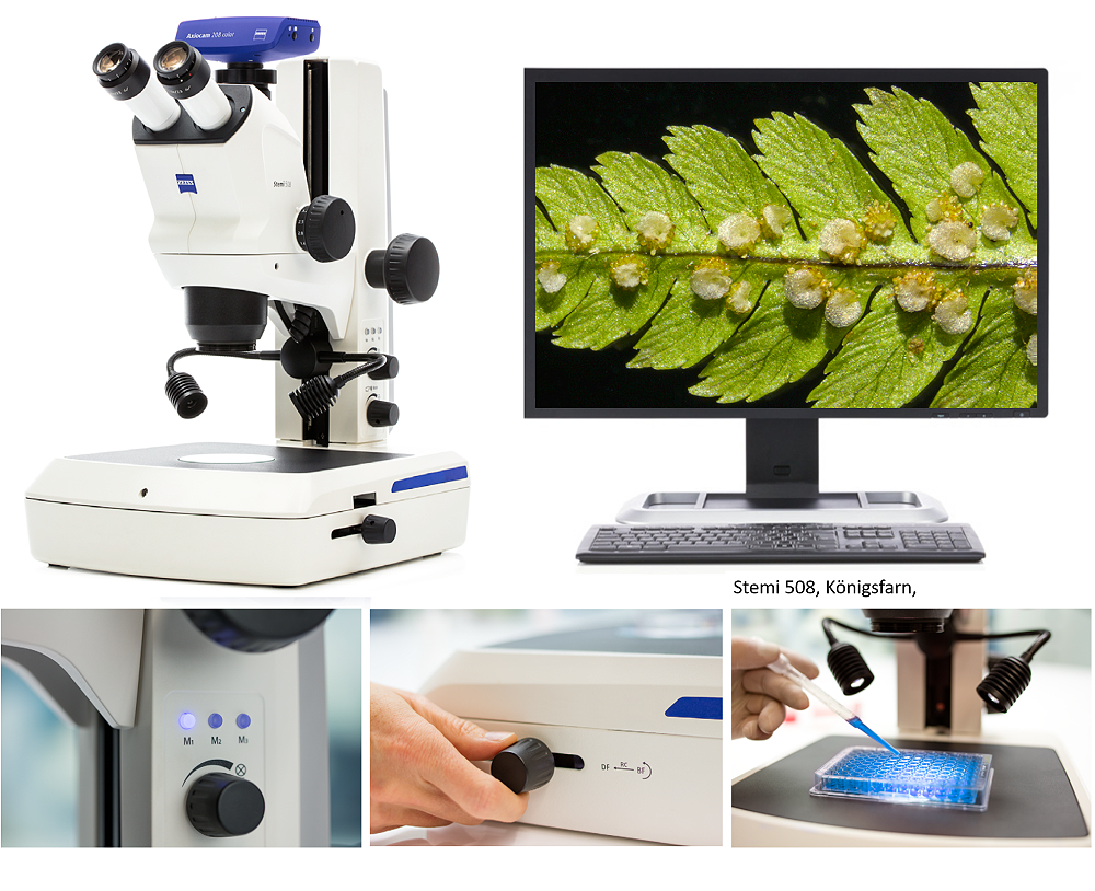
Choose between compact or flexible, stable boom stands, simple transmitted light or polarization contrast – tailor the configuration to your needs. Position your sample precisely with an additional rotating, sliding, or ball-joint table with polarization. Stemi 508 doc always comes with a C-Mount adapter for ZEISS Axiocam cameras, exchangeable for SLR or video cameras.
Microscope Stands
Stand M: Setup Memory for Rapid Recall
Save and quickly recall up to three lighting setups. The M LED stand comes equipped with built-in LED illumination. For transmitted light, choose between a mirror-based M LED unit or the flat bright-dark field transmitted light unit integrated into the stand base. Ideal for examining large samples or multiple Petri dishes simultaneously.
Stand K: Compact All-in-One Device
Transform your ZEISS Stemi 508 into a compact, easily deployable all-in-one device with the K stand. Options include the EDU stand for educational environments, LAB stand with mirror-based transmitted light, and MAT stand with incident light control and ESD features for quality inspection or small assembly work.
Stand N: Large Base for Large Samples
Benefit from a large stand base, a 350 or 450 mm high column, and a Stemi carrier for precise focusing on samples with large space requirements or height. Choose from sliding, ball, or rotating tables with polarization for tilting, shifting, or rotating your sample. The fiber optic cold light source provides daylight-like illumination, ideal for color-critical applications.
Boom Stands: Flexibility for Workspace and Sample Room
Use boom stands to observe large objects, locate and examine small samples in very large inspection areas, or quickly and flexibly pivot your microscope between different workstations. Large stands with boom arms simplify the movement of your Stemi 508 to any point in the workspace. The microscope always remains stable enough to comfortably observe small object details in stereo.
Illumination
Incident Light
Choose between the zoom- and height-adjustable K LED spot lamp for oblique and coaxial light with strong shadows, the self-supporting double spot LED with gooseneck for variable oblique light and clear shadow effects, and a segmentable LED ring light for shadow-free ring illumination.
Transmitted Light
Choose between the flat transmitted light base for bright- and dark-field illumination and the base with tiltable mirror for bright-field and dark-field illumination, as well as oblique light.
Applications:
Natural History Museums
Conserve, restore, digitize, and present collections in your museum.
Microscopy Solutions for Food Analysis
Detect ingredients, additives, and unwanted substances.
Microscopy Solutions for Embryo Quality Assessment
Criteria for "high-quality" embryos.
Microscopy Solutions for Oocyte Preparation and Quality
Assess the quality of oocytes.
Microscopy Solutions for Sample Preparation
For advanced studies such as genetic research.
Configurations (3)
Reviews
Select your country to view all the provider contacts:


