

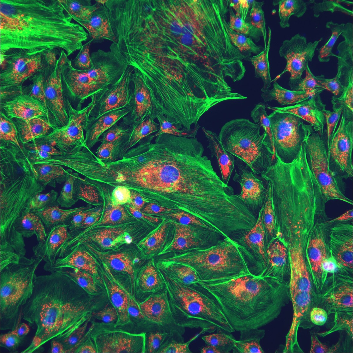
Fluorescence
© ETALUMA, INC.
The microscopic application of fluorescence is based on the phenomenon that certain substances (fluorophores) absorb light and then emit fluorescence when excited by light of a specific wavelength. This principle is utilized in fluorescence microscopy to gain detailed insights into biological structures and processes.
Details
Below, I explain the microscopic application of fluorescence and provide some examples of its use:
Microscopic application of fluorescence:
Immunofluorescence (IF): This technique uses fluorescently labeled antibodies to detect specific proteins or antigens in biological samples. The labeled antibodies bind to the target structures and enable visualization under a fluorescence microscope. IF is commonly used in cell biology and immunology to study the distribution of proteins in cells or detect the presence of antigens in tissue samples.
Fluorescence in situ hybridization (FISH): FISH is a technique used to locate specific DNA or RNA sequences in cells or tissues. Fluorescently labeled probes are used to hybridize to complementary sequences, allowing the identification of genomic regions, chromosomal abnormalities, or the study of gene expression.
Fluorescence Resonance Energy Transfer (FRET): FRET is a method to determine the proximity of molecules to each other. It relies on the transfer of energy between two fluorophores. When the molecules are in close proximity, the energy is transferred, leading to a decrease in fluorescence signal. FRET is used in protein research and the study of protein-protein interactions.
Application examples:
Investigation of cell structures: Fluorescence microscopy allows the examination of cellular components such as the cell nucleus, mitochondria, endoplasmic reticulum, and other organelles. This aids scientists in better understanding the functions and interactions within the cell.
Cancer research: Fluorescence microscopy is used in cancer research to characterize tumor structures, identify the presence of tumor markers, and investigate cell changes related to cancer.
Neurobiology: In neurobiology, fluorescence microscopy can be used to visualize neuronal connections, study synapses, and investigate neurodegenerative processes.
Pharmacology: Fluorescence microscopy is valuable in pharmacology to study the distribution and interactions of drugs in cells and tissues.
Biomedical research: Fluorescence microscopy is widely used in various other areas of biomedical research, such as the investigation of stem cells, infectious diseases, immune responses, and other biological processes.
The microscopic application of fluorescence has revolutionized biological research as it provides high-resolution images and information that traditional microscopy techniques cannot achieve.
Configurations
Reviews


